Best hair dye to use if you want dark black hair. Best hair dye out there for me nice and dark just how I like it.
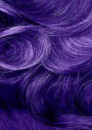
What colors mix well with purple?


MEET OUR BESTSELLERS
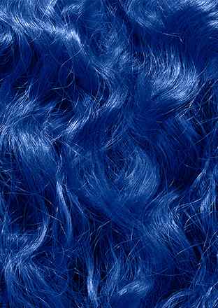
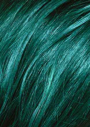
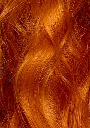
Hair dye colours to fall for

093 SHOCKING PINK


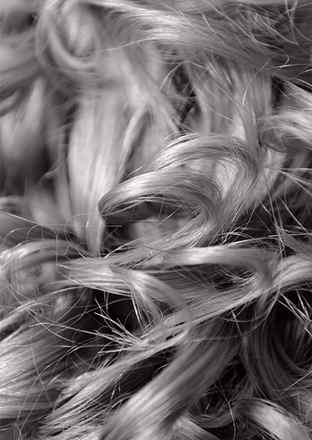
00B MAX BLONDE


030 MANGO TWIST

Inspiration
Don’t miss a thing about hair colour and hair dyes with LIVE.

Advice
GET READY TO FEEL INSPIRED BY OUR LIVE COLOUR ULTRA BRIGHTS

Advice
ALL ABOUT BLONDE HIGHLIGHTS: FRESH INSPIRATION FOR A CLASSIC LOOK
Bookshelf
NCBI Bookshelf. A service of the National Library of Medicine, National Institutes of Health.
StatPearls [Internet]. Treasure Island (FL): StatPearls Publishing; 2023 Jan-.
StatPearls [Internet].
Treasure Island (FL): StatPearls Publishing; 2023 Jan-.
Nishant Tripathi ; Amit Sapra .
Authors
Nishant Tripathi 1 ; Amit Sapra 2 .
Affiliations
1 University of Kentucky
2 Southern Illinois University School of Medicine
Last Update: August 14, 2023 .
The Gram staining is one of the most crucial staining techniques in microbiology. It gets its name from the Danish bacteriologist Hans Christian Gram who first introduced it in 1882, mainly to identify organisms causing pneumonia.[1] Often the first test performed, gram staining involves the use of crystal violet or methylene blue as the primary color.[2] The term for organisms that retain the primary color and appear purple-brown under a microscope is Gram-positive organisms. The organisms that do not take up primary stain appear red under a microscope and are Gram-negative organisms.
The first step in gram staining is the use of crystal violet dye for the slide’s initial staining. The next step, also known as fixing the dye, involves using iodine to form crystal violet- iodine complex to prevent easy removal of dye. Subsequently, a decolorizer, often solvent of ethanol and acetone, is used to remove the dye. The basic principle of gram staining involves the ability of the bacterial cell wall to retain the crystal violet dye during solvent treatment.[3] Gram-positive microorganisms have higher peptidoglycan content, whereas gram-negative organisms have higher lipid content.[4]
Initially, all bacteria take up crystal violet dye; however, with the use of solvent, the lipid layer from gram-negative organisms is dissolved. With the dissolution of the lipid layer, gram negatives lose the primary stain. In contrast, solvent dehydrates the gram-positive cell walls with the closure of pores preventing diffusion of violet-iodine complex, and thus, bacteria remain stained.[5] The length of decolorization is a critical step in gram staining as prolonged exposure to a decolorizing agent can remove all the stains from both types of bacteria.[6]
The final step in gram staining is to use basic fuchsin stain to give decolorized gram-negative bacteria pink color for easier identification. It is also known as counterstain. Some laboratories use safranin as a counterstain; however, basic fuchsin stains gram-negative organisms more intensely than safranin. Similarly, Hemophilus spp., Legionella app, and some anaerobic bacteria stain poorly with safranin.
Specimen Collection
Various clinical specimens can be used to perform Gram staining. Some of the commonly used specimens are sputum, blood, cerebrospinal fluid, ascitic fluid, synovial fluid, pleural fluid, and urine, etc. Swabs from nostrils, throat, rectum, wound, and cervix, etc. can also be used. The collection of specimens should always be in sterile containers.
Types of equipment needed for Gram staining include: